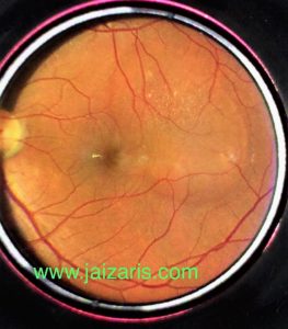
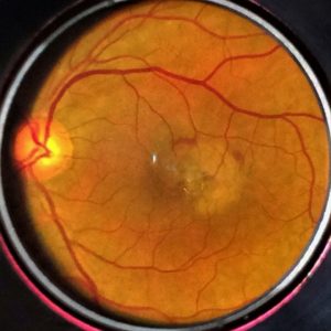
Branch Retinal Vein Occlusion
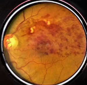
Due to block in a branch of retinal vein, lead to increase in blood back pressure leading to leakage of fluid and blood from vessels, which collects within the retina leading to reduced vision. Treatment options for this condition will the intravitreal injections of anti VEGF or Steroid, Sectoral laser.
If sectoral Laser is not done, there may be chances of Vitreous hemorrhage which may need surgery. Read more in section Floaters due to vitreous hemorrhage after BRVO.
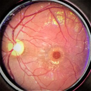
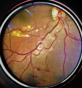
Branch Retinal Artery occlusion
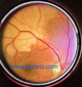
Branch Retinal Artery Occlusion is caused by infract which blocks Artery. And a part of retina function is lost leading to blurred sectoral field of vision.
These patients need systemic work up by physician or cardiologist to investigate about the place of origin of infract.
Central Retinal Vein Occlusion
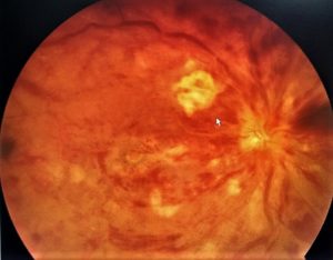
The major vein which leaves the eye is blocked leading to back pressure and bleeding and fluid leakage with the retina. Patient will notice it as sudden severe loss of vision.
Treatment options will be Intravitreal Anti VEGF or Steroid injection may be followed by PRP LASER to present Neo Vascular Glaucoma.
Central Retinal Artery Occlusion
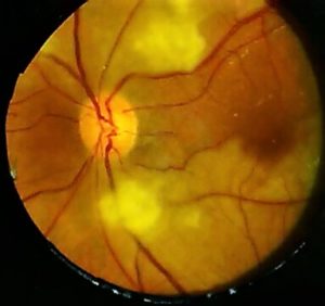
The central Retinal Artery which bring blood into the eye is blocked by infract at the entry to eye. This leads to Retinal Ischemia and Edema. Patient will experience sudden loss of significant vision. If patients present early within minutes or hours of onset of disease reducing intra ocular pressure by different technique may help in regaining part of vision.
These patients need to undergo systemic evaluation to know the origin of infract.
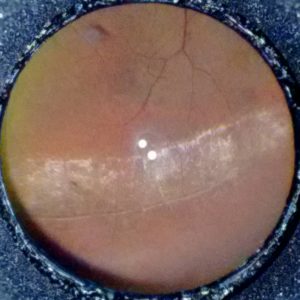
Central Serous Retinopathy
Collection of fluid below the macula, which leads to reduced vision in the centre of field.
Patient will notice vision is blurred in the center as a round area and beyond that vision is clear.
It may resolve or when indicated needs to investigate the source of fluid leakage by doing Fundus Fluorescein Angiography and laser the point of leak.
Macular Hole
This is usual age-related change, where the centre of retina develops Hole. This will lead to drop in vision for the patient.
Treatment of this disease is basically surgically management.
Lattice Degeneration
Among many of peripheral Retinal Changes, Lattice is the most common presentation. This is basically weak area of retina. Then lattice is associated with other risk factors, these may need Retina Barrage laser to prevent further enlargement and Retinal Detachment.
Retinal Tear
There can be hole Or Tear in the peripheral retina, which may extend to Retinal Detachment. These hole needs to be Barrage lasered.
Wet Age Related Macular Degeneration
With age the changes in the macula can be very significant causing fluid collection within macular area with or without bleed. Patients will have significant vision problem especially in the central visual field. These patients need further investigations and treatment. Treatment for some can be prolonged depending the severity of the disease.
Dry Age Related Macular Degeneration
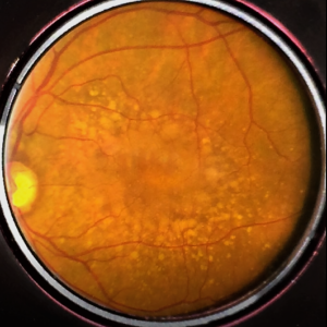
With age there will be changes in the Macular area, like pigmentary change, Drusen formation. These may affect vision in few. These needs yearly dilated pupil eye examination. Patient may or may not have vision problem.
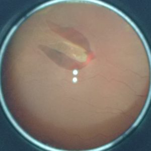
Valsalva Retinopathy
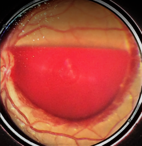
Sudden severe Sneezing or Stain, can lead to Blood collection between Retina and Vitreous gel. This lead to sudden loss of central vision.
On early presentation with Yag Hyaloidotomy, the blood will be displaced inferiorly resulting in regain of central vision.
Berlin Edema
When there is blunt injury to eye, Retina becomes edematous (whitening), which resolves over time. But there can be associated injury like choroidal tear, vitreous haemorrhage or late onset of Choroidal Neo vascular membrane. Any eye injury trivial or severe need regular follow up.